Baffled? This pathologist helps you make sense of your pathology report
By AMBER SMITH
Like many women who are diagnosed with breast cancer, Amanda Gilmore of Phoenix struggled with the new knowledge that her body contained cancerous cells that she could neither see nor feel.
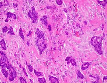
 Pathologists view cancer cells at the microscopic level, above, run tests and issue reports with a patient's test results but do not often interact directly with patients. An Upstate pathologist is taking a different approach and is happy to meet with patients to help explain what their pathology results mean.
She read her pathology reports, but she couldn’t understand them. She and her mother used the Internet for help with vocabulary, but it was still hard to put random words into context to know what they meant for her situation.
Then Gilmore’s surgeon, Kristine Keeney, MD, offered to bring Gilmore behind the scenes to meet with her pathologist.
A medical pathologist is a doctor who specializes in diagnosing and studying diseases using laboratory methods. He or she may supervise tests on blood and other bodily fluids and tissue, such as the breast tissue removed during Gilmore’s biopsy.
Pathologist Rohin Mehta, MD, says pathologists are the doctors’ doctors. Typically their work is in the background, with little if any direct patient contact. That’s changing in some areas of the country. Lowell General Hospital in Massachusetts launched a pathologist-patient consultation program last year called Cancer Pathology 101.
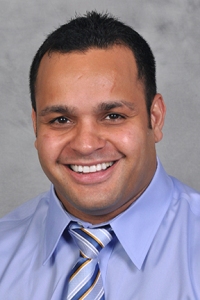
 Rohin Mehta, MD
At Upstate, Mehta has begun a more modest undertaking. He offers to meet one-on-one with breast cancer patients who want help understanding the pathology of their cancer.
“I can see how scared they are, and this can give them some solace,” he says.
For Gilmore, 38, seeing images of what the cancer cells looked like in comparison to her healthy cells, with Mehta’s explanation, made her realize her situation was not so dire.
“He definitely reassured us,” she recalls. “Dr. Mehta was personable and very easy to talk to. After I met with him, I felt much better about all of it.
“It made me feel like breast cancer was no longer a foreign concept.”
Gilmore was diagnosed after a biopsy in October. Keeney operated to remove the mass in her breast in November. Then Gilmore went through radiation therapy. She
finished in January and because “my cancer showed it was reactive to estrogen,” she takes a medication to block the estrogen.
Mehta enjoys the patient interaction.
Not everyone wants to read their pathology report or see what their cancer looks like under a microscope. And many patients are satisfied with answers they can get from their primary doctor. For those with a lingering desire to know more, Mehta tries to oblige.
“I figure it’s the right thing to do,” he says. “This lets patients kind of envision what they’re fighting against.”
A few terms from a breast cancer pathology report
1 o’clock excision — refers to the geographic location of a tumor or tissue sample that was removed, if you envision the face of a clock on the breast; 1 o’clock would be above and slightly to the right of the nipple
gross description — the pathologist’s overall description of the tissue; it may be a few words or a whole paragraph
mitotic rate — tells how quickly the cancer cells are dividing
tumor focality — the number of tumors; if there are many, it’s multifocal
macrometastasis and micrometastasis — when cancer has spread to a lymph node, it will be called micro if it measures less than 2 millimeters and macro if it encompasses the whole lymph node

 This article appears in the spring 2019 issue of Cancer Care magazine. Click here to hear an interview with Rohin Mehta, MD, about how to understand a pathology report.


